
Magnetic Resonance Imaging (MRI)
Have a doctor’s order for an imaging study?
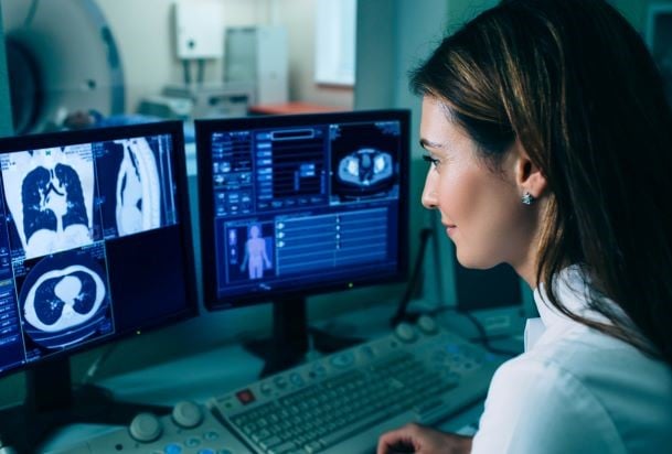
Powerful Imaging Technology
Magnetic resonance imaging, or MRI, is a non-invasive procedure that uses powerful magnets and radio waves to create pictures of the body, often used in medical imaging for soft tissues.
MRI is ideal to help diagnose many types of injuries and conditions because of the ability to tailor the exam to the particular medical question being asked. Unlike conventional X-rays and computed tomography (CT) which use radiation, MRI imaging is based on the magnetic properties of atoms. Some common uses for MRI include:
Imaging knee & shoulder injuries involving muscles, ligaments & tendons
Imaging the brain, spinal cord & nerves
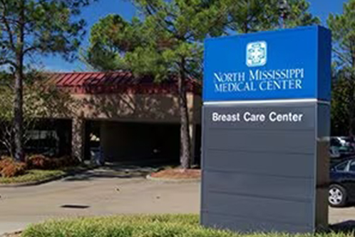
Breast cancer screening for those at high risk
When frequent imaging is needed, MRI can be used instead of CT reduce radiation exposure.
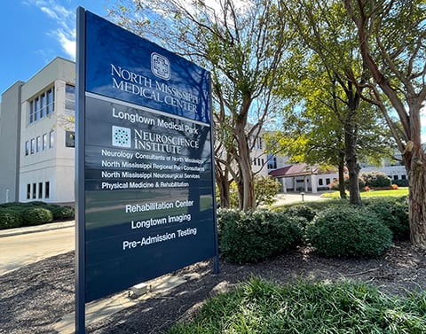
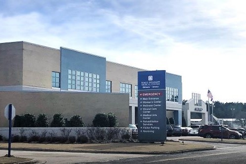
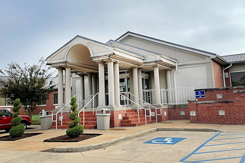
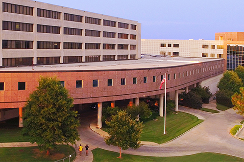
MRI is available with a physician’s order at NMHS locations across north Mississippi and northwest Alabama including:
The Imaging Center, Tupelo
Longtown Medical Imaging, Tupelo
Medical Imaging at Barnes Crossing, Tupelo
North Mississippi Medical Center-Tupelo
North Mississippi Medical Center Gilmore-Amory
North Mississippi Medical Center-Iuka
North Mississippi Medical Center-West Point
North Mississippi Medical Center-Hamilton, Alabama
MRI machines are large, tube-shaped magnets. The patient lies down a padded table for the imaging study.
MRI exams average 30-60 minutes.
You will need to lie still to avoid blurry images.
Depending on your study, you may need a contrast agent by IV or injection.
The MRI machine will not touch you during the exam.
There can be loud clicking and beeping noises.
If you are claustrophobic, talk to your doctor about special accommodations.
Related Locations
Imaging Awards
Mammography
Accredited by the American College of Radiology
Computed Tomography (CT)
Accredited by the American College of Radiology
Magnetic Resonance Imaging (MRI)
Accredited by the American College of Radiology
Radiology Department
Accredited by the State of Mississippi
Related Resources
View AllNurse Link® is a free telephone triage and health information service provided by North Mississippi Health Services. Using computerized medical protocols, nurses direct callers to the most appropriate medical treatment.
Cancer Screening
Radiology plays an important part in screening and early detection for breast cancer and lung cancer. Breast cancer screening starts with mammography. Lung cancer screening uses low dose CT.
Cancer Screening
Radiology plays an important part in screening and early detection for breast cancer and lung cancer. Breast cancer screening starts with mammography. Lung cancer screening uses low dose CT.