Summary
In this episode of We’re All Heart, Dr. Barry Bertolet interviews Dr. Murray Estess, a non-invasive cardiologist. Dr. Estess explains the role of echocardiography in diagnosing heart conditions, emphasizing the advantages of 3D echo technology and how advanced imaging improves patient outcomes.
We're All Heart: Episode Three
Podcast Transcript
Introduction: Life is about the moments that make your heart tick, and we’re here to keep it ticking strong. I’m Dr. Barry Bertolet, and this is We're All Heart, where we dive into the latest cardiac care with the experts who live it every day.
From breakthrough procedures to the most cutting-edge treatments, we're putting heart health front and center.
We're All Heart is brought to you by North Mississippi Health Services in partnership with Cardiology Associates of North Mississippi. Let's get to it.
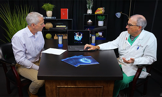
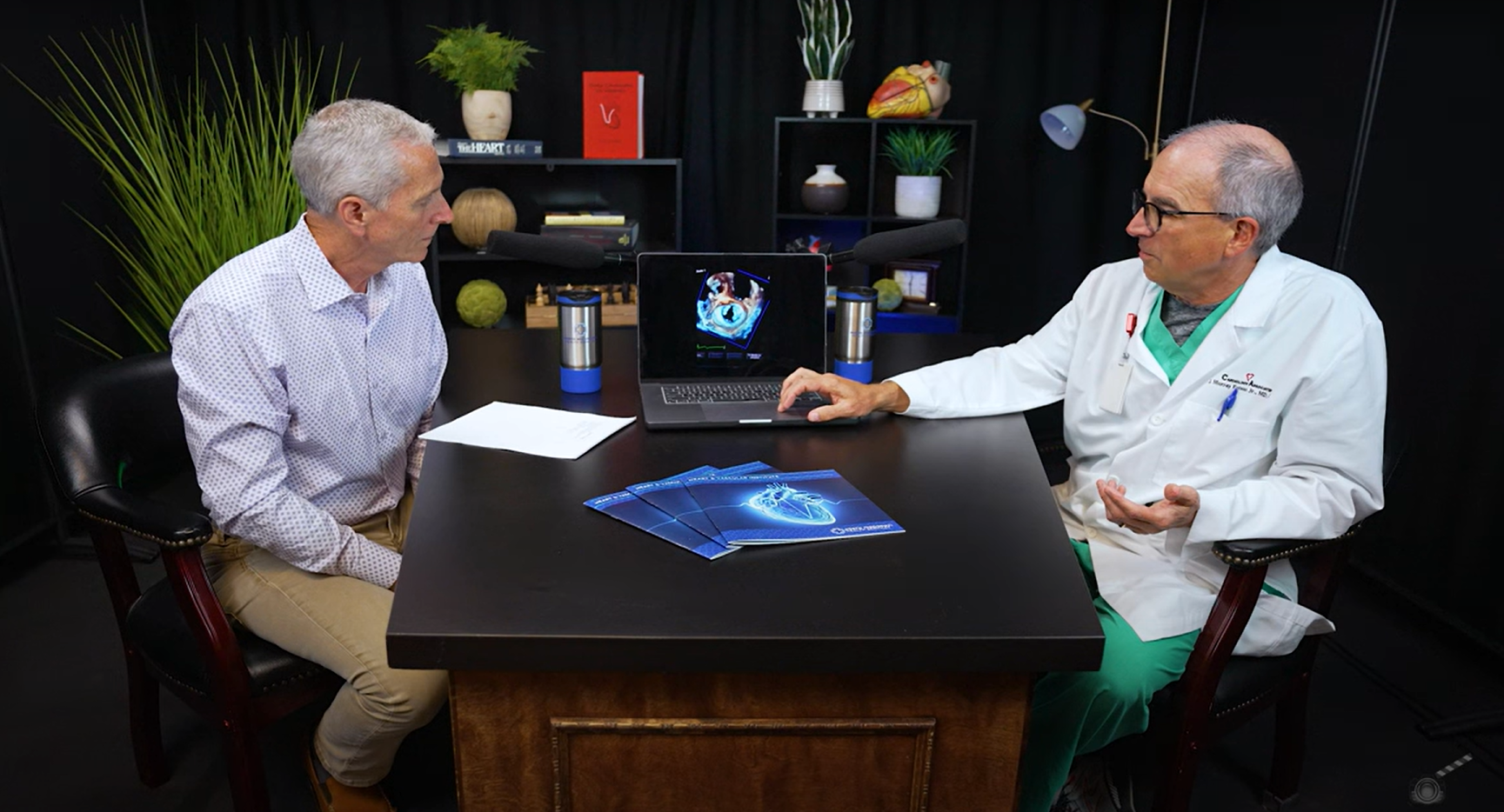
Dr. Barry Bertolet: I'm Dr. Barry Bertolet and I'm here with my partner Dr. Murray Estess, who is an echocardiography expert. And he's going to be talking to us today about 3D Echo.
So I'd like to welcome Dr. Estess. Murray, tell us a little bit about your background and your training and what brought you to Tupelo.
Dr. Murray Estess: Thank you Dr. Bertolet. Well, I started out as a pharmacist. I graduated pharmacy school, then I went to medical school. I did my residency at the University of Virginia in Charlottesville, Virginia. And then I did my cardiology fellowship at the Cleveland Clinic and I've been here now for 23 years.
Dr. Bertolet: I didn't hear any Mississippi there. So what got you back to Mississippi?
Dr. Estess: Well, as it just turns out, my dad was playing golf with one of the ENTs here and he happened to tell Dr. Calhoun that I was training. And Cardiology Associates called me and recruited me here and I've been here ever since.
Dr. Bertolet: He needed a job, so. That's great. And we're so happy to have you here. And you've been here now how long?
Dr. Estess: 23 years.
Dr. Bertolet: 23 years. That's quite a long time. So tell me what your specialties within cardiology might be.
Dr. Estess: Okay, so I am a what you call non-invasive cardiologist. So I do transesophageal echocardiograms, I do regular echocardiograms. I do nuclear stress testing, stress echocardiography, and I see patients in the clinic as well.
Dr. Bertolet: So you're doing imaging on people where you're not sticking stuff in them?
Dr. Estess: That's right, that's right. Sometimes we have people swallow a tube - called a transesophageal echo and we take pictures. But that's about as far as I go.
Dr. Bertolet: And that imaging that you're doing that will detect blockages in blood vessels?
Dr. Estess: The imaging that we do when we do stress testing will detect blockages in blood vessels. We can look at blood flow in the heart using PET scan and CT scans and we can actually detect blockages that way.
Dr. Bertolet: And then the echocardiogram, what does that do?
Dr. Estess: So the echocardiogram is an ultrasound test. It's where we pass ultrasound waves through the chest. We are able to see those ultrasound waves come back to the echo probe. And that gives us an image of the heart.
We can see the valves, we can see the function of the heart, we can see blood flow. It's very much like the same technology that you use to see a baby when a baby's in the womb. (Continued)
We're All Heart
Join Dr. Bertolet for more episodes featuring local experts in all areas of heart health. We'll learn about the cutting-edge treatments offered right here in north Mississippi - often before the rest of the state or even nation. New episodes drop on YouTube or anywhere you get your podcasts every other week.
Dr. Bertolet: So I was watching the Hunt for Red October this past weekend and the submarine used these sonars and then that's how they were picking up where something else was. Is that sort of what we're talking about when we're talking about an echo?
Dr. Estess: That's right. A sound wave is sent out, it bounces off an object and comes back and the echo machine can interpret those sound waves, make pictures, and we can see those sound waves come back and form what the heart actually looks like inside.
Dr. Bertolet: And does that hurt?
Dr. Estess: It doesn't hurt at all.
Dr. Bertolet: Nice. And how do the sound waves go in? What do you do to deliver the sound wave?
Dr. Estess: The echo machine is equipped with a probe. The probe sends out sound waves from the probe. We push it against the chest, sound waves go in, harmlessly, bounce off the heart and come back.
Dr. Bertolet: And how quickly do you get information back from that?
Dr. Estess: Immediately.
Dr. Bertolet: Immediate information. And when we do an echocardiogram, what are some of the, I'm gonna say the disease processes that we might be looking for there?
Dr. Estess: So sometimes the heart's weak, we can detect weaknesses in the heart muscle itself. We can look at the valves. There are four valves in the heart and we can see all four valves.
We can see whether those valves are diseased, whether they might be narrowed where blood flow is not going through adequately or in cases where blood flow is actually leaking from those valves that aren't closing properly.
Dr. Bertolet: So if someone were to have a problem with heart failure symptoms, then they may want to get an echocardiogram.
Dr. Estess: That's right. That would be one of the main reasons you would get one.
Dr. Bertolet: And then if there was a concern about heart valve disease, that would be another good reason to have a echocardiogram.
Dr. Estess: Definitely.
Dr. Bertolet: How many, when we think about that echo that's on the chest, how many dimensions does a normal echo show?
Dr. Estess: Well, the normal echo is just like your camera at home. Your normal camera is gonna give you two dimensions.
But we live in a 3D world, we actually see things in three dimensions. You may have watched a movie that showed 3D but it's not really 3D it's only an illusion of 3D. It's very hard to see in three dimensions.
Dr. Bertolet: So if I were a patient and you were examining my heart, I would probably want you to see all the heart, like in all three dimensions as opposed to just in two dimensions is like maybe like a piece of paper or something like that. Is there anything better than just two dimensional echo?
Dr. Estess: Well, we rely on 2D echo quite a lot, but now we have the ability using echo to do three dimensional echocardiography.
Dr. Bertolet: Wow.
Dr. Estess: So that's something fairly new, but we've been doing it here probably about 10 years, I would say.
Dr. Bertolet: So would that be like if mama's having a new baby and she pays the extra little money and she gets the baby's face before it's born, that's sort of that 3D echo. Is that sort of what we're looking at here too?
Dr. Estess: Very, very similar. Exactly the same technology. We can use that for the heart and see things in greater detail that we were not able to appreciate before.
Dr. Bertolet: And when we're talking about that 3D echo, I mean, so you're able to see the heart as it looks, I'm gonna use the word anatomically in the body. So you're able to look at a body part or a part of the heart non-invasively in a way to look at that in really great detail.
Dr. Estess: It's not exactly the way the heart looks, but it's pretty close.
Dr. Bertolet: So if you were a surgeon or an interventionalist and you needed information to plan a surgery, you would have a three-dimensional model, so to speak, of the heart before you went in and did something?
Dr. Estess: Yes. We can take pictures before the surgery, show the surgeon exactly what kind of valvular pathology he's likely to see when he opens up the heart to look at that valve. We can look at the echocardiogram after the procedure is done and see what has been done. And we can get a three dimensional representation before the patient is closed up. We can look at that in detail and see if everything is satisfactory as it's actually functioning after surgery.
Dr. Bertolet: That's nice. That's a big advantage. And I understand that you brought some examples for us to look at today.
Dr. Estess: I did, I brought some pictures here. So this is an example of a normal mitral valve. And you can see that the mitral valve, the way it opens and closes inside the body without having to open the patient up, you can actually look and you can see exactly what that valve looks like.
Dr. Bertolet: So the mitral valve is located where?
Dr. Estess: The mitral valve is located between the left atrium and the left ventricle, the two main chambers of the heart. The left ventricle is the one that does all the work.
I have some views of the mitral valve here. This is actually a patient who had a cleft mitral valve. Normally the mitral valve should be the same on both sides with a valve here and a valve leaflet here.
Dr. Bertolet: So I see that hole there.
Dr. Estess: We see a tear in the leaflet. And this is a congenital deformation of the mitral valve and that we were able to really appreciate that in three dimensions.
Dr. Bertolet: So that's a, that's basically a birth defect of the mitral valve that you've seen.
Dr. Estess: That's right.
Dr. Bertolet: So with that birth defect that's there, what would be a symptom that a person would have with that birth defect?
Dr. Estess: So a person might have shortness of breath when they're doing activity.
The eventually may have enlargement of both the left atrium and the left ventricle and could develop heart failure from that.
Dr. Bertolet: So with that hole in the mitral valve, would that cause leakiness? Would every time the heart contracted would blood slash back into the left atrium in the lungs?
Dr. Estess: That's right, that's right.
Dr. Bertolet: So that's what would cause the shortness of breath. Because you have fluid sloshing back in the lungs.
Dr. Estess: Yes. Yes. And so sometimes we might fix that valve with a mechanical valve. And in this case you can see that there's a mechanical valve right here. The valve is open and closing, and so we can get a better appreciation of how that valve works. Or we might have a valve like this where this is a bioprosthetic valve or a valve made out of cow or pig pericardial tissue.
Dr. Bertolet: So if somebody had to have open heart surgery and had a valve put in, that's what it would look like.
Dr. Estess: That's right. And these are three dimensional images of what that valve would look like when it's open and closing. And this is a normal looking valve. And so in cases where we have and we can see that this valve is not opening well and that valve is a diseased prosthetic valve that's not working right.
Dr. Bertolet: I can clearly see that - that's not my specialty, but I can even make that diagnosis and I hope that you can too, seeing that one valve was opening quite well and this one is not opening well.
So what would the surgeon do with information such as that?
Dr. Estess: So when a surgeon sees that, or a cardiologist might see that, they might send that patient back to the surgeon to say, hey, we need to put in a new valve, or we need to do something to fix this. Now we actually have the technology where a valve like that, we can actually go in with a catheter and on that catheter is mounted a new valve and we can actually slide that valve into that old valve without having to open the patient back up and do surgery.
Dr. Bertolet: So a minimally invasive heart valve surgery?
Dr. Estess: Yes, that would be called a valve-in-valve transcatheter valve replacement.
Dr. Bertolet: So I will just tell the audience, join us in a future podcast because I'll be talking to Dr. David Talton about minimally invasive heart valve surgery. So stay tuned for that in the future
Dr. Estess: Sometimes we, we see unusual things in three-dimensional echo. For example, this patient here had a tumor that was in the heart, and you can see that the tumor is in this top chamber of the heart and you can get an appreciation. This is the left ventricle here in the bottom, and this is the left atrium and this is the tumor that's in the heart. And so that gives us a visual appreciation of what might be going on.
Dr. Bertolet: Now you don't hear about tumors in the heart that often. How often does a tumor occur? And then if you found that, what would you do with that?
Dr. Estess: Tumors are very unusual in the heart, but in a case like this, this is big enough that that tumor has to come out.
So that would help when we can and we image that further, we can help the surgeon know exactly where he needs to go to get that tumor.
Dr. Bertolet: Well, so that would lead to a resection of the tumor and removal of the tumor and hopefully a cure for the patient.
Dr. Estess: Yes.
Dr. Bertolet: Just starting with a simple ultrasound.
Dr. Estess: Yes. Most tumors of the heart are benign, but some aren't.
Dr. Bertolet: Right. Well, this has been absolutely fascinating to look at here. And so I guess a question that I would have is that how many hospitals around us are even doing three dimensional echo?
Dr. Estess: There are probably not many hospitals around us that are doing three dimensional echo.
Usually only centers that have the machines that are capable. One, it takes a certain type of machine to be capable. Most of the newer echo machines that are sold are capable, but most of the hospitals that probably not bought the software.
Dr. Bertolet: So again, maybe some technology that North Mississippi has here for a decade, it sounds like - that other hospitals in our surrounding area have not adopted this technology yet.
But you've convinced me. I'm convinced that this would make a huge difference for our patients here
Final question. We're leading off with True North, and so that is a campaign of North Mississippi Health Services. And so I would like to ask you, what is your true north, what are the reasons why you're here?
What do you think that you would like to do in your practice here? What would you like to see as outcomes for your patients?
Dr. Estess: Well, we'd like to see the best outcomes we can have, and I think we've got a great center here.
We've got great support from the medical community. We've got good primary care physicians. We have good cardiologists. We also have great cardiovascular surgeons that support us. And I think having a solid team.
I always like to say that the reason teams win national championships is because they have a deep bench. And I think that we have a deep bench here and I think that we have a chance to provide great service for our patients and we really strive for the best outcomes that we can have.
Dr. Bertolet: That's wonderful. That's a great segue.
So thank you for joining us and if you want to see more of our deep bench from North Mississippi Medical Center come join me on the next podcast.
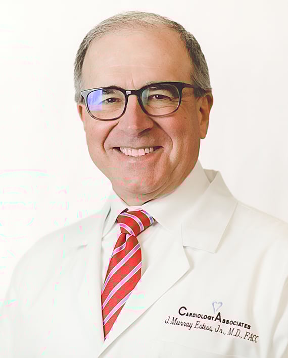
Murray Estess, MD
Subscribe
Follow us on Spotify or Apple Podcasts or subscribe to our YouTube channel so you never miss an episode!
Related Links
Topics
- Managing Your Health
- We're All Heart
Share
If you're over 35, schedule a heart screening to identify your risk of heart disease. Request an appointment online or call 1-800-THE DESK (1-800-843-3375).

Subscribe to Our Newsletter
Like this content and want to get more? Sign up for True North, the health and wellness newsletter from North Mississippi Health Services!
Subscribe to Our Newsletter
Like this content and want to get more? Sign up for True North, the health and wellness newsletter from North Mississippi Health Services!

Nurse Link®
Not sure if you need Urgent Care or the ER? Call 1-800-882-6274 anytime to speak directly to a registered nurse and get immediate answers. Using computerized medical protocols, nurses direct callers to the most appropriate treatment. Our nurses are available 24 hours per day, seven days per week.